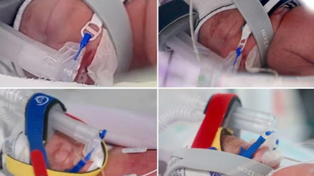
An Australian mother recently made global headlines after naturally conceiving and giving birth to identical quadruplet girls, an extraordinary event estimated to occur in roughly one in 11 million to one in 64 million pregnancies. All four infants developed from a single fertilized egg that split three distinct times in early development, sharing identical genetic makeup. While media coverage often frames these rare births as pure feel-good miracles, the underlying reproductive biology and high-risk obstetric management required to bring four identical fetuses to term present one of the most intense technical challenges in modern maternal-fetal medicine.
The odds of naturally conceiving identical quadruplets are so infinitesimal that most practicing obstetricians will never encounter a single case across their entire careers. To put the phenomenon into perspective, non-identical higher-order multiples usually stem from fertility treatments, where multiple eggs are fertilized or multiple embryos are transferred. Spontaneous monozygotic quadruplets happen entirely by chance, independent of maternal age, family history, or reproductive technology.
Understanding how a single zygote splits into four distinct embryos requires breaking down the timeline of early cellular division.
The Cellular Mechanics Behind the Split
In a standard pregnancy, a single sperm fertilizes a single egg to form a zygote, which then begins rapid cell division as it travels down the fallopian tube. Monozygotic, or identical, multiples occur when this early cluster of cells spontaneously divides into separate entities.
For quadruplets to form identically, the initial fertilized egg must undergo a sequential series of divisions within the first week after fertilization.
Day One to Day Three
The first split occurs during the cleavage stage. If the cellular mass divides completely at this point, two identical embryos are created. Each will go on to develop its own outer membrane (chorion) and inner membrane (amnion). This is known as a dichorionic diamniotic structure.
Day Four to Day Eight
If one or both of those newly divided cellular masses split a second time during the blastocyst stage, additional distinct embryos form. Depending on the exact timing of these secondary splits, the embryos may end up sharing a placenta or sharing an amniotic sac.
The Third Division
To reach four identical embryos, a third split must take place. The structural configuration of the resulting pregnancies varies depending on whether the splits happen early or late. The most common survivable configuration for identical quadruplets involves two placentas, where two sets of identical twins each share a placental mass, though far more precarious arrangements can occur.
If cellular division happens after day 13 post-fertilization, the separation remains incomplete, resulting in conjoined twins rather than separate individuals.
The High-Stakes Physics of a Monochorionic Gestation
Carrying multiple fetuses creates extreme physiological demands on the human body, but the real medical danger stems from shared vascular systems. When fetuses share a placenta, they share a blood supply.
In monochorionic pregnancies, microscopic vascular connections form within the shared placental tissue, connecting the circulatory systems of the fetuses. This structure creates significant operational risk for the clinical team managing the pregnancy.
Twin-to-Twin Transfusion Syndrome
One of the most dangerous complications in a shared placental environment is Twin-to-Twin Transfusion Syndrome (TTTS). This condition occurs when blood flows disproportionately from one twin (the donor) to another (the recipient) through interconnecting placental blood vessels.
The donor fetus receives inadequate blood volume, leading to slowed growth, poor kidney function, and dangerously low amniotic fluid levels. Meanwhile, the recipient fetus receives an excess of blood, overloading its cardiovascular system and potentially causing heart failure and excessive amniotic fluid accumulation. In a quadruplet pregnancy where multiple fetuses share placental tissue, tracking these circulatory imbalances requires weekly high-resolution Doppler ultrasound imaging.
Twin Anemia-Polycythemia Sequence
A more subtle variant is Twin Anemia-Polycythemia Sequence (TAPS), where tiny vascular connections allow a slow transfer of red blood cells from one fetus to another without the dramatic amniotic fluid shifts seen in TTTS. One fetus becomes severely anemic while the other develops hyperviscous, thick blood. Unchecked, TAPS can result in neurological injury or intrauterine demise for both fetuses.
Selective Fetal Growth Restriction
When four fetuses share limited placental real estate, the distribution of nutrient-rich tissue is rarely equal. One fetus may end up with an offset, marginal umbilical cord insertion that provides only a fraction of the necessary surface area for nutrient exchange. This leads to Selective Fetal Growth Restriction (sFGR), where one or more quadruplets lag far behind their siblings in weight and organ development.
| Complication Risk | Single Pregnancy | Identical Quadruplet Pregnancy |
|---|---|---|
| Premature Labor (<32 weeks) | Extremely Low (<2%) | Near Certain (95%+) |
| Preeclampsia | 3% - 5% | Up to 60% |
| Placental Vascular Complications | 0% | High (Requires weekly monitoring) |
| Gestational Diabetes | 6% - 10% | 40% - 50% |
Managing the Extreme Physiology of Higher-Order Multiples
The human uterus is evolutionarily optimized to carry a single fetus weighing between six and eight pounds. Carrying four fetuses simultaneously forces the maternal cardiovascular, respiratory, and metabolic systems to operate at their absolute upper limit.
Maternal blood volume increases by roughly 40% to 50% during a single pregnancy. In a higher-order multiple pregnancy, that expansion can exceed 100%. The maternal heart must pump twice as much blood per minute to supply the uterus, kidneys, and peripheral tissue, causing a dramatic increase in cardiac output and resting heart rate.
The physical volume of four growing fetuses, along with four amniotic sacs and multiple placentas, places massive mechanical pressure on internal organs. The maternal diaphragm is displaced upward early in the second trimester, significantly reducing lung capacity and causing chronic shortness of breath.
The risk of severe preeclampsia—a dangerous condition characterized by high blood pressure and protein leakage in the urine—skyrockets in quadruplet pregnancies due to the excessive amount of placental tissue releasing inflammatory factors into the mother's bloodstream.
To manage these cascading physical demands, maternal-fetal specialists deploy strict monitoring protocols starting early in the second trimester.
- Bi-weekly or weekly ultrasound examinations to monitor individual fetal growth rates, amniotic fluid volumes, and placental blood flow dynamics.
- Continuous blood pressure and renal function screening to detect early signs of preeclampsia or HELLP syndrome.
- Proactive cervical length checks to assess the structural integrity of the cervix under heavy mechanical weight.
- Targeted nutritional therapy designed to support massive cellular expansion without provoking severe gestational diabetes.
The Delivery Strategy and Neonatal Intensive Care Operations
A full-term human pregnancy lasts approximately 40 weeks. For identical quadruplets, reaching 40 weeks is biologically impossible and clinically dangerous. The average gestational age for quadruplets at delivery is around 28 to 30 weeks.
Holding a quadruplet pregnancy past 32 weeks carries significant risks of uterine rupture, placental abruption, and stillbirth due to placental insufficiency. Consequently, delivery is planned weeks in advance via scheduled Cesarean section, usually executed by a massive multidisciplinary medical team.
A quadruplet delivery is not a simple surgical procedure; it is a synchronized trauma-level operation requiring weeks of simulations.
The Surgical Protocol
Two lead obstetric surgeons perform the Cesarean section, accompanied by scrub nurses, circulating nurses, and anesthesiologists dedicated solely to maintaining maternal stability. Because the uterus is severely distended, the risk of postpartum hemorrhage—massive bleeding caused by failure of the uterine muscle to contract after delivery—is extremely high. Surgical teams must have rapid-infusion blood products on standby before making the initial incision.
The Neonatal Resuscitation Teams
Every infant requires their own dedicated resuscitation station and medical team inside the operating room. For four infants, this means four separate teams composed of:
- A neonatologist or pediatric hospitalist.
- A neonatal intensive care unit (NICU) registered nurse.
- A respiratory therapist specialized in premature airway management.
As each baby is delivered, they are immediately passed to their designated team, stabilized, placed in temperature-controlled thermal wrap to prevent rapid heat loss, intubated or placed on continuous positive airway pressure (CPAP) if necessary, and transported to the NICU in specialized isolettes.
The Long Road Through the NICU
Delivering four premature infants safely is only the first phase of the clinical intervention. Micro-premature infants born before 30 weeks gestation face months of intensive care as their underdeveloped organ systems mature.
The primary hurdle for infants born at 28 to 30 weeks is respiratory distress syndrome. Their immature lungs lack sufficient pulmonary surfactant, a lipid-protein substance that keeps the tiny air sacs (alveoli) from collapsing upon exhalation. Neonatal teams must administer synthetic surfactant directly into the lungs through an endotracheal tube shortly after birth.
Beyond breathing support, premature quadruplets face several critical physiological hurdles in the NICU.
Intraventricular Hemorrhage
The delicate blood vessels in the brain of a premature infant are highly susceptible to rupturing during changes in blood pressure, leading to bleeding into the brain's ventricular system. Specialized neurosonography is used to monitor for intraventricular hemorrhage during the first critical weeks of life.
Necrotizing Enterocolitis
The gastrointestinal tract of a 28-week infant is unequipped to digest standard nutrients. Necrotizing enterocolitis (NEC) is a devastating inflammatory disease where bacteria invade the wall of the intestine, potentially causing tissue necrosis and bowel perforation. Supplying mother's milk or pasteurized donor human milk alongside specialized intravenous nutrition (total parenteral nutrition) is vital for minimizing this risk.
Retinopathy of Prematurity
Because the blood vessels serving the retina are among the last to fully develop in a fetus, premature exposure to fluctuating oxygen levels can cause abnormal vessel growth. This condition, known as retinopathy of Prematurity (ROP), can lead to retinal detachment and vision loss if not tracked and treated with laser therapy or specialized injections.
The extraordinary birth of identical quadruplets in Australia represents a remarkable convergence of statistical anomaly, cellular mechanics, and advanced maternal-fetal intervention. While public attention focuses on the biological wonder of four identical faces, the true achievement lies in the complex clinical navigation that allows a mother and four critically premature infants to survive one of the rarest medical events in human reproduction.